Imaging
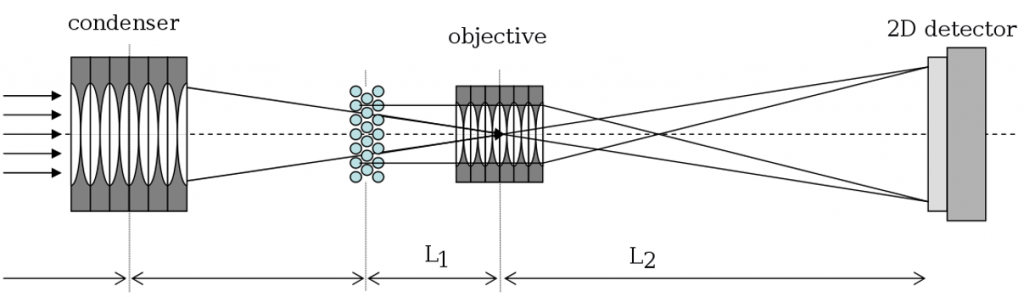
A common application for refractive lenses from RXOPTICS is x-ray imaging in transmission mode. Usually, illumination of the object from behind is achieved by a prefocusing lens (condenser) in order to adjust the beam size to the sample (Figure 1, cf. the section on focusing). The image is formed by an objective lens with a small focal length which allows a large magnification L2 / L1. A large magnification relieves the requirements on the resolution of the x-ray detector.
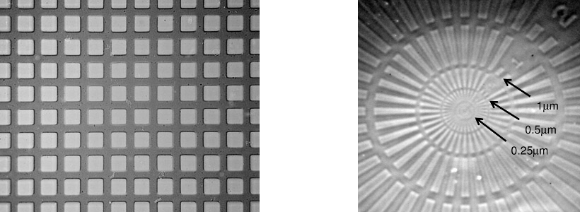
Figure 2 (left) shows a Nickel mesh with a period of 12.7 μm that was imaged at 12 keV by 91 beryllium 2D lenses with R = 0.2 mm and a magnification factor L2 / L1 = 10 onto a high resolution film (from Lengeler et al. (2005): Refractive x-ray lenses). The distance of the objective CRL from the sample was L1 = 546 mm, yielding an image distance of L2 = 5460 mm. It is obvious that the image is practically free of aberrations.
Figure 2 (right) shows a Ta Siemens star that is imaged at 46 keV by Al 2D lenses with a magnification factor L2 / L1 of 13 (private communication with M. DiMichiel, M. Scheel, A. Snigirev, I. Snigireva, ESRF). This CRL was made up of 60 lenses with R = 0.05 mm and 321 lenses with R = 0.2 mm. The distance of the objective CRL from the sample was L1 = 906 mm, resulting in an image distance of L2 = 11,778 mm. The condenser CRL, consisting of 125 Al 2D lenses with R = 0.2 mm, was placed 2.4 m from the sample and, thus, produced a virtual focal spot inside the objective lens (Figure 1).
Note that for CRLs containing such a large number of lenses it is important to take into account thick lens effects; in particular, sample and image distance have to be understood with respect to the primary and the secondary principal plane, respectively.
Diffraction
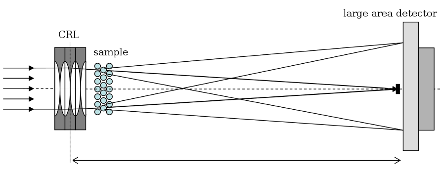
Another major application of refractive lenses from RXOPTICS is x-ray diffraction. Apart from the usual configuration employing a collimated beam, high-resolution x-ray diffraction can be performed by removing the objective lens in the configuration in Figure 1 and placing a large area detector in the back focal plane of the condenser (Figure 3). By Fresnel theory, the detector measures the Fraunhofer diffraction pattern.
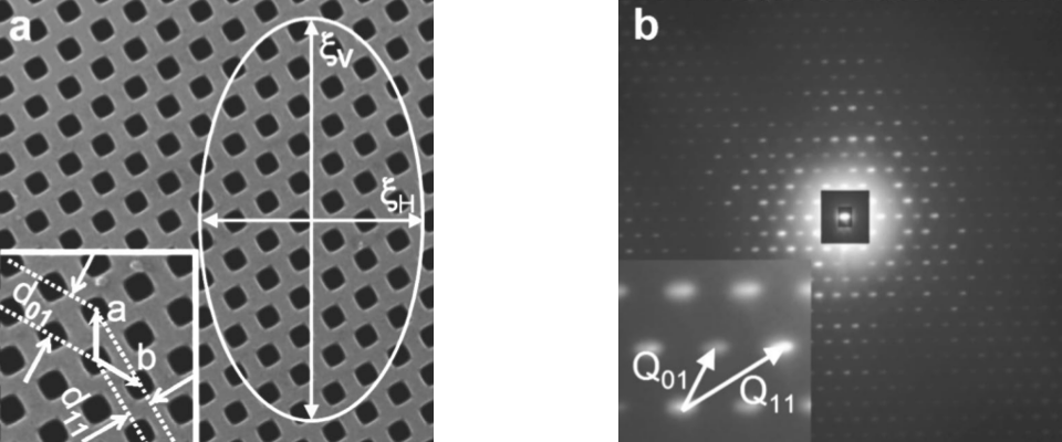
This particularly compact experimental setup has been used by M. Drakopoulos et al. to investigate a two-dimensional photonic crystal fabricated into a 150 μm thick Si wafer (Figure 4 and Drakopoulos et al.: X-ray high-resolution diffraction using refractive lenses). The CRL consisted of 112 Al 2D lenses with R = 0.2 mm and a focal length of 1334 mm at 28 keV. CRL and sample were located 58 m from the undulator source of beamline ID18F at ESRF. The two vectors a and b defining the hexgonal grid of the photonic crystal have a length of 4.2 μm. The two easily resolved lattice vectors Q01 and Q11 in reciprocal space have a length of 1.75 × 10⁻³ nm-1 and 2.99 × 10⁻³ nm-1, respectively (Figure 4, right). In this configuration, resolution is not limited by the divergence of the x-ray source but by its small angular size of about 1 μrad; the achieved resolution was of the order 10-4 nm-1.