
Focusing
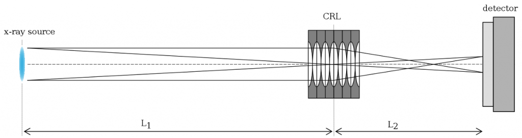
Focusing of the x-ray beam is one of the major applications of refractive x-ray lenses from RXOPTICS. Focusing maps the primary x-ray source to a focal spot, the so-called secondary source (Figure 1).
This is done, for instance, to adjust the beam to the sample size and to increase the photon flux on the sample in x-ray diffraction, absorption, and fluorescence experiments. The gain in intensity, that is, the ratio of the intensities in focal spots of the same size produced by the CRL on the one hand and by a pinhole on the other hand, can be of the order of 104 and above. For instance, a CRL consisting of 52 ideal beryllium 2D lenses with R = 0.2 mm maps a source at 15 keV with a size of 300 μm × 15 μm (FWHM) at a distance L1 = 40 m to a secondary source at a distance L2 = 1330 mm with a size of 10.0 µm × 0.5 µm (FWHM); the gain is 4.7×10⁴. While 2D lenses are used for rotionally symmetric focusing, astigmatic focusing or focusing in only one direction is possible by employing 1D lenses (cf. the section on preconditioning).
Figure 2 compares the intensity profiles of the beam of the ID18 at ESRF, Grenoble, at 14.41 keV with and without a CRL in the vertical and in the horizontal direction (private communication with A. Chumakov, ESRF). Here, the source is mapped by a CRL made up of 39 beryllium 2D lenses with R = 1.5 mm. The secondary source is well fitted by a Gaussian profile (solid red lines) and hence shows very low background in the wings. The Gaussian intensity profile is a result of the parabolic form of the lenses.

The absence of ringing, that is, significant parts of the intensity residing in oscillations apart from the main focal spot, is an important advantage of parabolic lenses over other x-ray optics and plays a crucial role in many experiments.
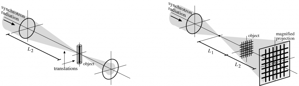
Focusing and microscopy
In some techniques, like scanning transmission x-ray microscopy and ptychography (scanning x-ray diffraction microscopy), the secondary source is scanned over the sample (Figure 3, left). A small secondary source may also be used in x-ray phase-contrast microscopy (Figure 3, right). Placing the object close to the focus yields a strong magnification M = L2 / L1. The smaller the focus, the sharper is the image.
Collimation
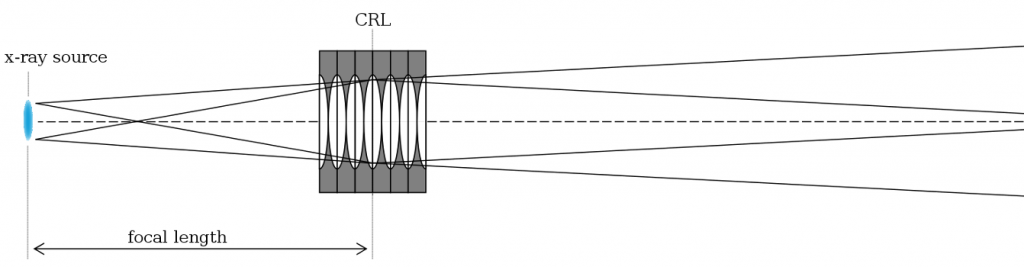
Collimation of divergent x-ray sources, which is particularly important in diffraction experiments, is in a sense the inverse operation to focusing. For collimation, the CRL is placed at a distance from the source which (roughly) equals its focal length; the angular size of the source, that is, the ratio of its lateral extent and its distance to the CRL, then translates into a residual divergence of the collimated beam (Figure 4).